
How to Choose the Best Bone Biopsy Needle in 2026?
Choosing the best bone biopsy needle in 2026 is not a catalog exercise. It is a clinical decision shaped by the lesion, the route, imaging guidance, and the tissue needed for diagnosis. A dense pelvic target may call for a different approach than a small, fragile vertebral lesion. The needle must reach the intended site while supporting adequate sampling and limiting avoidable trauma. Small details matter: cortical thickness, lesion depth, access angle, and whether the sample must preserve tissue architecture.
Interventional radiologist Dr. Michael J. Wallace is an established specialist in image-guided procedures. No verified quotation from him on bone biopsy needle selection was supplied, so attributing one would be misleading. A useful editorial principle is: “Match the needle to the target, the route, and the tissue requirement.” It is a guiding idea, not a sourced quotation.
This guide examines needle design, gauge, length, cutting mechanism, compatibility with imaging, and practical handling. It also considers specimen quality and the value of following the manufacturer’s instructions and local clinical protocols. No single model is best for every case. That is easy to forget when product specifications look convincing. Clinicians should weigh evidence, training, and patient-specific anatomy, then discuss uncertain choices with the relevant specialist. The right selection supports a careful procedure; it cannot replace clinical judgment.
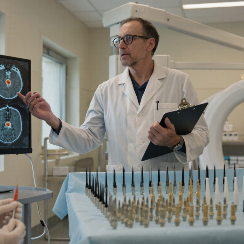
Understand the Purpose and Clinical Setting of Bone Biopsy
Bone biopsy needle selection begins with a clear question: what must the sample help diagnose? A suspected infection may require material for culture, while a possible tumor may need enough tissue for microscopic and molecular analysis. The intended test can affect how the team plans the procedure and handles each specimen.
The target matters just as much. A dense lesion near a joint is different from a soft, accessible area in a long bone. Imaging helps clinicians assess its size, position, and nearby structures. The patient’s anatomy, bleeding risk, and ability to remain still also shape the plan. Small details matter.
In a hospital, a radiologist or other trained clinician may coordinate with pathology before the procedure. They can discuss whether core tissue, aspirated material, or both are needed. A needle that reaches the target safely but yields too little tissue may not answer the clinical question. That trade-off is easy to underestimate.
There is no single best needle for every setting. In practice, the decision is not always tidy; imaging, sample requirements, and patient comfort may point in different directions. Clinicians should weigh these factors against local expertise and the specific diagnostic goal.
How to Choose the Best Bone Biopsy Needle in 2026? — Understand the Purpose and Clinical Setting of Bone Biopsy
| Clinical Setting | Primary Diagnostic Purpose | Typical Sampling Approach | Needle Selection Considerations | Guidance and Targeting | Key Planning Points |
|---|---|---|---|---|---|
| Suspected bone metastasis | Identify tumor type and obtain tissue for histology and, when needed, molecular testing. | Image-guided percutaneous core biopsy; multiple cores may be collected when safe and appropriate. | Choose a core needle and gauge that balance tissue volume with safe access. The lesion’s density, location, and surrounding structures affect the choice. | CT or ultrasound guidance may be used depending on lesion visibility and access. Target viable tissue and avoid areas that appear necrotic when possible. | Review imaging and any planned treatment tests beforehand. Confirm specimen handling requirements with pathology, especially if molecular studies are anticipated. |
| Suspected primary bone sarcoma | Establish a diagnosis while preserving options for definitive surgical treatment. | Image-guided core biopsy planned by, or in consultation with, the orthopedic oncology team. | A core biopsy approach is commonly considered; the route and needle path matter as much as the needle itself. | Use cross-sectional imaging to plan a path that avoids unnecessary contamination of nearby compartments and structures. | Coordinate biopsy planning with the treating tumor team before the procedure. The biopsy tract may need to be removed during definitive surgery. |
| Bone marrow disorder | Assess marrow cells and architecture for conditions such as hematologic malignancy or unexplained cytopenias. | Bone marrow aspiration together with a trephine core biopsy is commonly performed, usually from the posterior iliac crest in adults. | A marrow trephine needle is selected to obtain an adequate core; aspiration and core sampling provide complementary information. | Usually performed using anatomical landmarks, with imaging considered when anatomy or access is difficult. | An aspirate alone may not provide enough information about marrow architecture. Follow local protocols for specimen preparation and allocation. |
| Suspected bone infection | Identify the infecting organism and assess tissue for infection or other causes of the lesion. | Image-guided sampling of bone or an associated collection; specimens may be sent for microbiology and histopathology. | Choose an approach that can safely reach the target and provide enough material for the requested tests. | CT or other suitable imaging guidance can help target the affected area while avoiding nearby structures. | Arrange separate specimen handling for culture and histology. Do not place the microbiology specimen in formalin; coordinate collection and transport with the laboratory. |
| Vertebral bone lesion | Diagnose a vertebral lesion, including suspected tumor or infection. | Image-guided percutaneous core biopsy; a transpedicular route is commonly considered when anatomically suitable. | Needle type and gauge depend on the target, bone quality, access route, and proximity to neural and vascular structures. | CT guidance is frequently used to plan and monitor access to vertebral targets. | Review cross-sectional imaging and assess bleeding risk, stability, and the safest trajectory. Coordinate specimen allocation with the clinical question. |
| Small, densely sclerotic, or difficult-to-access lesion | Obtain a diagnostic sample from a target where limited tissue yield or access may increase the risk of a nondiagnostic result. | Individualized image-guided sampling; the target and approach are selected after reviewing imaging and clinical context. | Consider whether a needle can penetrate the lesion and retrieve adequate tissue without creating unacceptable procedural risk. | Use the imaging modality that best depicts the lesion and a safe access route; targeting may be adjusted based on lesion characteristics. | Discuss the expected yield and alternatives with the relevant specialists. A nondiagnostic biopsy may require repeat sampling or another diagnostic approach. |
Selection principle: There is no single best bone biopsy needle for every case. Needle design and gauge should be chosen according to the diagnostic goal, lesion characteristics, anatomical access, imaging guidance, and specimen requirements. Biopsy planning should be individualized by the treating clinical team.
Related Posts
-
China Top Bone Biopsy Needle Manufacturers and Suppliers
-
2025 Top Bone Biopsy Needle Techniques for Effective Diagnosis and Treatment
-
How to Choose the Right Bone Biopsy Needle for Accurate Diagnosis?
-
How to Choose the Right Bone Biopsy Needle for Your Medical Needs
-
What is a Bone Marrow Biopsy Kit and How is it Used?
-
10 Best Bone Marrow Needle Options for Efficient Biopsy Procedure?