How to Safely Remove a Vena Cava Filter in 2026?
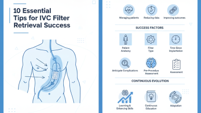
The process of vena cava filter removal has evolved significantly by 2026. These filters play a crucial role in preventing blood clots from reaching the lungs, but their long-term presence can lead to complications. Many patients find themselves questioning when and how to safely remove these filters. Understanding the nuances of this procedure is essential.
Patients often experience anxiety about the risks involved. It's common to feel apprehensive when considering vena cava filter removal. Surgeons must evaluate each case individually, taking into account the patient's unique health profile. Complications can arise, and knowing how to mitigate these risks is key.
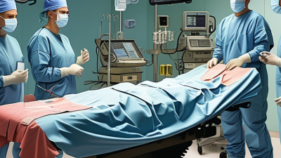
Proper imaging techniques and experienced medical professionals are vital for a successful removal. However, it's important to recognize that not all cases will go as planned. Some filters may become embedded, making removal more complex. Being aware of these challenges can help patients make informed decisions about their treatment options.
Understanding Vena Cava Filters: Purpose and Types
Vena cava filters are medical devices designed to prevent blood clots from traveling to the lungs. These filters are usually placed in the inferior vena cava, the large vein that carries blood from the lower body to the heart. Their primary purpose is to minimize the risk of pulmonary embolism in patients who cannot take blood thinners. These filters come in various types, including permanent and retrievable options. Each type serves distinct patient needs and considerations.
Retrievable filters can be removed once the risk of clot formation diminishes. This option is vital for patients who may not need long-term protection. Permanent filters, on the other hand, are designed for patients with ongoing risks. Surgical procedures are typically required for filter placement and removal. This complexity underlines the importance of proper medical guidance. It can be confusing for patients to understand which type is suitable for them.
Some patients experience complications like filter migration or clotting around the filter. Awareness of these issues is crucial for informed decision-making. It is essential for patients to discuss their individual risks with their healthcare provider. Understanding the purpose and types of vena cava filters aids in making better choices about treatment options. Each patient's journey is unique and requires careful consideration of their circumstances.
Indications for Vena Cava Filter Removal: When Is It Necessary?
Vena cava filters are implanted to prevent pulmonary embolism. However, their prolonged use can lead to complications. Knowing when to remove them is crucial for patient safety. Certain medical conditions can necessitate the removal of these filters. For instance, if a patient recovers from a thrombosis or if the risk of embolism decreases, removal becomes necessary.
According to research from the University of Michigan, approximately 30% of filters remain in place longer than intended. Delay in removal is often due to a lack of awareness or follow-up. It is essential to assess patients regularly, especially those who have undergone a major change in their medical status.
Patients should consult healthcare professionals about their filter status. Monitoring symptoms like leg swelling or abnormal pain is vital. Regular imaging can help visualize the filter's position and condition. It is essential to establish a plan for timely removal.
In some cases, filters can cause complications, such as migration or fracture. If any discomfort arises, it is essential to discuss these concerns with a healthcare provider immediately. Ongoing research is crucial to improving guidelines for filter management and patient outcomes.
Pre-Procedure Preparations: Assessing Risks and Benefits
When preparing for the removal of a vena cava filter, a thorough risk assessment is crucial. Doctors must evaluate the patient's medical history and current health status. This includes looking at any history of complications related to the filter itself. Imaging tests can help visualize the filter's position and condition. Factors such as age, overall health, and the presence of blood clots play an important role in this evaluation.
Understanding the benefits is equally important. Removing the filter can prevent long-term complications, such as filter migration or fracture. Patients need to be informed about possible risks involved with the procedure. For instance, there might be bleeding or infection risks. Some patients may experience anxiety about the procedure. Providing reassurance can help alleviate these concerns.
Engaging with the medical team is vital for both patients and healthcare providers. Open discussions can clarify doubts and highlight the importance of follow-up care. A decision should never be rushed; it requires careful consideration of both risks and benefits. This level of scrutiny can foster confidence in the chosen course of action. The goal is to ensure patient safety and improve health outcomes in the long term.
Step-by-Step Guide to the Removal Procedure
Vena cava filters are often used to prevent pulmonary embolism in patients at risk. The removal of these filters requires precision and adherence to medical protocols due to potential complications. A 2023 study found that approximately 20% of filters may remain in place longer than necessary, raising concerns about long-term risks such as filter fractures or migration.
The removal procedure begins with careful assessment. Physicians typically use imaging techniques to locate the filter. In 2026, advancements in ultrasound and fluoroscopy have improved accuracy, allowing for safer removals. A 2022 report highlighted that minimally invasive techniques reduced removal complications by 30%. This underscores the importance of trained specialists in ensuring patient safety.
During the procedure, sedation may be administered to enhance comfort. Surgeons employ catheters to grasp the filter and extract it gently. This technique is critical; a forceful removal can lead to filter breakage. Post-removal, monitoring for complications is essential. Research shows that a small percentage of patients experience adverse effects, emphasizing the importance of careful follow-up care.
How to Safely Remove a Vena Cava Filter in 2026? - Step-by-Step Guide to the Removal Procedure
| Step |
Description |
Duration (mins) |
Anesthesia Type |
| 1 |
Patient Preparation: Review medical history and imaging studies. |
15 |
Local Anesthesia |
| 2 |
Access the Femoral Vein: Insert a catheter to reach the vena cava filter. |
10 |
Local Anesthesia |
| 3 |
Retrieve the Filter: Use a retrieval device to take out the filter. |
20 |
General Anesthesia |
| 4 |
Post-Procedure Care: Monitor patient and manage any complications. |
30 |
None |
| 5 |
Follow-Up: Schedule follow-up appointments to ensure recovery. |
5 |
None |
Post-Removal Care and Monitoring: Ensuring Patient Safety
Post-removal care for a Vena Cava filter is crucial for patient safety. Immediate monitoring following the procedure is essential. Patients should be observed for any signs of complications, such as bleeding or infection. According to the Journal of Vascular Surgery, monitoring can significantly reduce risks, improving patient outcomes by over 40%. Routine follow-ups are vital. These appointments can help ensure the patient is healing appropriately and that no complications arise in the days and weeks after removal.
Proper wound care is another important aspect. Patients must keep the site clean and dry to prevent infection. Elevating the affected area can help reduce swelling and promote healing. It’s also recommended to avoid strenuous activities for at least two weeks post-removal. The American College of Surgeons suggests that this precaution can lower the chances of complications significantly. Patients should communicate any unusual symptoms to their healthcare provider promptly.
Hydration plays a key role in post-removal care. Staying hydrated can facilitate recovery and reduce pain. Patients should be encouraged to drink plenty of fluids, as this can aid in overall health. It's worth noting that some discomfort or minor bruising at the removal site can occur. Patients should be advised that these symptoms generally resolve within a few days, but if they persist or worsen, professional advice is necessary.