What Is a Hysterosalpingogram Catheter Used For?

What Is a Hysterosalpingogram Catheter Used For?

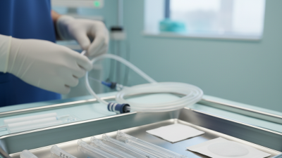
A hysterosalpingogram catheter is a narrow medical device used during hysterosalpingography, or HSG. It gently delivers contrast material through the cervix and into the uterus. X-ray imaging then shows the uterine cavity and whether the fallopian tubes are open. The catheter is not designed to treat infertility. It helps clinicians investigate possible structural causes.

The World Health Organization reported in 2023 that infertility affects approximately one in six people worldwide. WHO Director-General Dr. Tedros Adhanom Ghebreyesus stated, “Infertility affects millions of people of reproductive age worldwide.” That reality gives this procedure practical importance. A catheter may help reveal blocked tubes, uterine abnormalities, or unexpected cavity changes. Small details matter, including cervical anatomy, balloon pressure, contrast volume, and catheter stability.

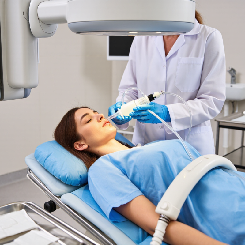
The American Society for Reproductive Medicine identifies tubal assessment as an important part of selected infertility evaluations. The Radiological Society of North America also describes HSG as an imaging examination for evaluating the uterus and fallopian tubes. In a clinical room, the process is precise: the patient lies beneath an X-ray unit, a speculum provides access, and the hysterosalpingogram catheter is positioned before contrast enters slowly.

Results are useful, but not absolute. Tubal spasm can sometimes resemble blockage. Discomfort can also affect the examination. Therefore, interpretation should combine imaging, symptoms, medical history, and professional judgment. This is where the explanation becomes less tidy. A catheter provides evidence, not certainty. Its value depends on careful placement, appropriate imaging, and qualified clinical review.
What Is a Hysterosalpingogram Catheter Used For?

What Is a Hysterosalpingogram Catheter?

What Is a Hysterosalpingogram Catheter?

A hysterosalpingogram catheter is a thin, sterile tube used during an HSG examination. A trained gynecologist or radiologist gently places it through the cervix and into the uterine cavity. Its main purpose is controlled delivery of contrast medium. The contrast outlines the uterus and travels toward the fallopian tubes while X-ray images are taken.

The catheter often includes a soft tip or small balloon that helps create a temporary seal. This seal can reduce contrast leakage through the cervix. It also helps the clinician direct the fluid without using excessive pressure. The catheter is not an imaging device. It is the access route for the examination.

HSG can help assess the shape of the uterine cavity and whether the fallopian tubes appear open. The American Society for Reproductive Medicine recognizes tubal evaluation as an important part of infertility assessment. The World Health Organization reported in 2023 that infertility affects about one in six people worldwide. That figure explains why this relatively small procedure remains clinically relevant.

Some patients feel cramping when contrast enters the uterus. Others feel only brief pressure. Experience varies, and the process is not always as comfortable as patient guides suggest. Proper consent, infection screening, pregnancy exclusion, and careful pressure control matter. Catheter placement should stop if severe pain or unusual resistance occurs.

How the Catheter Supports Hysterosalpingography

What Is a Hysterosalpingogram Catheter Used For?

How the Catheter Supports Hysterosalpingography

A hysterosalpingogram catheter helps deliver contrast material through the cervix during an HSG examination. This X-ray procedure evaluates the uterine cavity and checks whether the fallopian tubes are open. A trained clinician gently places the catheter after reviewing the patient’s history, consent, and possible risks.

The catheter creates a controlled pathway for the contrast fluid. Some designs use a small balloon, while others rely on a soft tip or cervical seal. These features can help limit leakage and improve the clarity of the images. The clinician slowly introduces the fluid while monitoring its movement on an X-ray screen. That movement outlines the uterus and may show contrast passing through the tubes.

The experience varies. Mild cramping or pressure is common, but discomfort should not be ignored. Catheter placement can feel awkward, and the first attempt is not always perfect. Cervical position, prior procedures, and individual anatomy may affect the technique. Sterile handling matters because infection, although uncommon, remains a possible complication. Patients should ask about pain relief and report fever, heavy bleeding, or severe pain afterward. The catheter supports the examination, but it does not diagnose infertility by itself. A qualified clinician must interpret the images alongside other medical findings.

What Is a Hysterosalpingogram Catheter Used For?

A hysterosalpingogram catheter is inserted through the cervix to create a controlled pathway for contrast material. It helps maintain a seal, supports steady contrast delivery, and allows X-ray imaging of the uterine cavity and fallopian tubes.

The chart shows the main procedural roles of the catheter and the approximate sequence in which they support hysterosalpingography. Actual timing varies according to patient anatomy, imaging technique, and clinical practice.

When a Hysterosalpingogram Catheter Is Used

When a Hysterosalpingogram Catheter Is Used

A hysterosalpingogram catheter is used during an HSG examination. It gently enters the cervix and delivers contrast material into the uterus. X-ray images then show the uterine cavity and fallopian tubes. Doctors commonly recommend HSG during infertility evaluations, especially when blocked tubes are suspected. The World Health Organization reported that infertility affects about one in six people worldwide (WHO, 2023). Tubal factors remain an important part of that investigation. The catheter is diagnostic, not a fertility treatment.

HSG is usually scheduled after menstrual bleeding ends but before ovulation. This timing reduces the chance of testing during an early pregnancy. It may also help investigate repeated miscarriages, unusual uterine shapes, or previous pelvic infections. The American Society for Reproductive Medicine lists HSG as a standard method for assessing tubal patency (ASRM, 2021). Still, the test is not perfect. Temporary tubal spasm can sometimes resemble blockage. A clear image cannot guarantee pregnancy.

Tips: Ask whether pregnancy testing is needed before the procedure. Tell the clinician about allergies, pelvic pain, or possible infection. Take only recommended pain relief. Mild cramping and light spotting may occur afterward. Severe pain, fever, or heavy bleeding needs prompt medical advice. Personal experiences vary, and that uncertainty deserves honest discussion.

What Is a Hysterosalpingogram Catheter Used For? - When a Hysterosalpingogram Catheter Is Used

Clinical Dimension How the Hysterosalpingogram Catheter Is Used Typical Information Obtained
Primary purpose A thin catheter is placed through the cervix into the uterine cavity to deliver a water-soluble or oil-based contrast medium during an X-ray examination. The examination outlines the inside of the uterus and shows whether the fallopian tubes allow contrast to pass through them.
Evaluation of infertility The catheter is used during a hysterosalpingogram when tubal blockage or an abnormal uterine cavity may be contributing to difficulty becoming pregnant. Results may help identify blocked or narrowed tubes, an unusually shaped uterine cavity, or other findings that require further evaluation.
Assessment of fallopian-tube patency Contrast is injected slowly through the catheter while images are taken as the medium enters the uterus and moves toward the tubes. Free passage of contrast into the pelvic cavity generally suggests that the fallopian tubes are patent. Lack of passage may suggest obstruction, although spasm can sometimes affect the appearance.
Assessment of the uterine cavity The catheter provides a route for contrast to fill and outline the uterine cavity. Imaging may show filling defects or contour changes associated with conditions such as endometrial polyps, submucosal fibroids, adhesions, or certain uterine shape variations.
Evaluation after selected procedures In appropriate cases, an HSG catheter may be used for imaging after tubal surgery or a sterilization procedure, according to the treating clinician’s protocol. The study can help assess whether the intended tubal result has occurred or whether the tubes remain open.
Timing in the menstrual cycle HSG is commonly scheduled after menstrual bleeding has ended and before ovulation, often during the first half of the cycle. The exact timing varies by clinical protocol and cycle length. Scheduling is intended to reduce the possibility of performing the examination during an unrecognized pregnancy and to provide a clear view of the uterine cavity.
Basic procedure sequence The patient is positioned for a pelvic examination, a speculum is inserted, the cervix is cleaned, and the catheter is gently introduced. Contrast is then injected while fluoroscopic X-ray images are obtained. The images show the pathway of contrast through the uterus and, when open, through the fallopian tubes into the pelvic cavity.
Expected sensations Insertion of the catheter and injection of contrast may cause temporary cramping or pressure. The intensity varies between individuals. Mild cramping and light spotting can occur after the examination. Persistent or severe symptoms should be reported to a healthcare professional.
Important safety considerations The procedure should not be performed when pregnancy is suspected. Active pelvic infection, unexplained significant bleeding, or a known allergy to the planned contrast medium should be discussed before the examination. Medical history, allergies, symptoms of infection, and pregnancy possibility are reviewed before scheduling or performing the test.
What the catheter does not provide The catheter is a delivery and access device; it does not independently diagnose infertility or treat every cause of reduced fertility. HSG findings are interpreted together with medical history, pelvic examination, laboratory testing, ultrasound, and other fertility assessments when needed.
Clinical note: A hysterosalpingogram is an imaging procedure performed by trained healthcare professionals. The choice of catheter, contrast medium, timing, and follow-up depends on the patient’s medical history and the facility’s clinical protocol.

What Happens During Catheter Placement

During a hysterosalpingogram, catheter placement begins after pregnancy has been excluded. The patient lies on an X-ray table with knees bent. A speculum gently opens the vagina, allowing the clinician to see the cervix clearly. Antiseptic solution cleans the area. Sometimes, a small instrument steadies the cervix. It can feel sharp briefly.

The clinician then guides a thin catheter through the cervical opening. Its tip rests inside the uterus. A small balloon or soft seal may hold it in place. Placement should be controlled, not forced. Mild cramping is common when the catheter enters or contrast begins flowing. Clear communication matters. Tell the team if pain becomes sudden, severe, or persistent.

Fluoroscopy shows whether contrast travels through the uterine cavity and fallopian tubes. The American Society for Reproductive Medicine identifies HSG as a standard test for tubal patency. The World Health Organization’s 2023 infertility report estimates that about 17.5% of adults experience infertility during their lives. That makes accurate placement clinically important. Published reviews referenced in radiology guidance report pelvic infection rates below 1% in appropriately screened patients, although rates vary. The procedure is usually brief. Still, placement is not always effortless. Cervical position, anxiety, prior procedures, or discomfort can change the experience. A small technical adjustment may be needed. Safety is careful work, not a guarantee.

Safety Considerations and Possible Discomfort

What Is a Hysterosalpingogram Catheter Used For?

A hysterosalpingogram catheter helps deliver contrast material through the cervix during an X-ray examination. The contrast outlines the uterus and fallopian tubes, showing whether the tubes are open.

A clinician usually places the thin, sterile catheter after confirming that pregnancy is not present. ACOG advises discussing radiation exposure, allergies, and infection risks before imaging.

The dose is generally low, but “low” does not mean zero. Timing also matters, usually after menstruation and before ovulation.

Discomfort Is Common

Discomfort is common, although intensity varies greatly. Patients may feel pressure, brief cramping, or a sharp sensation when contrast enters the uterus. Some experience light spotting afterward.

According to the American College of Radiology and Radiological Society of North America, pelvic infection occurs in fewer than 1% of examinations.

Contrast reactions are uncommon, but staff should know about previous allergies.

Pain medicine may help, only if the clinician approves it.

Slow breathing can reduce tension, though it cannot remove every sensation. That distinction matters.

Seek medical advice for fever, worsening pelvic pain, heavy bleeding, faintness, or unusual discharge.

These symptoms are uncommon but should not be ignored.

A catheter can be positioned carefully and still feel uncomfortable. Personal pain reports are not perfectly predictable, and clinical studies cannot capture every patient’s experience. Clear instructions, sterile technique, and immediate communication remain essential safety measures.