Top 10 Questions About Vena Cava Filter Removal

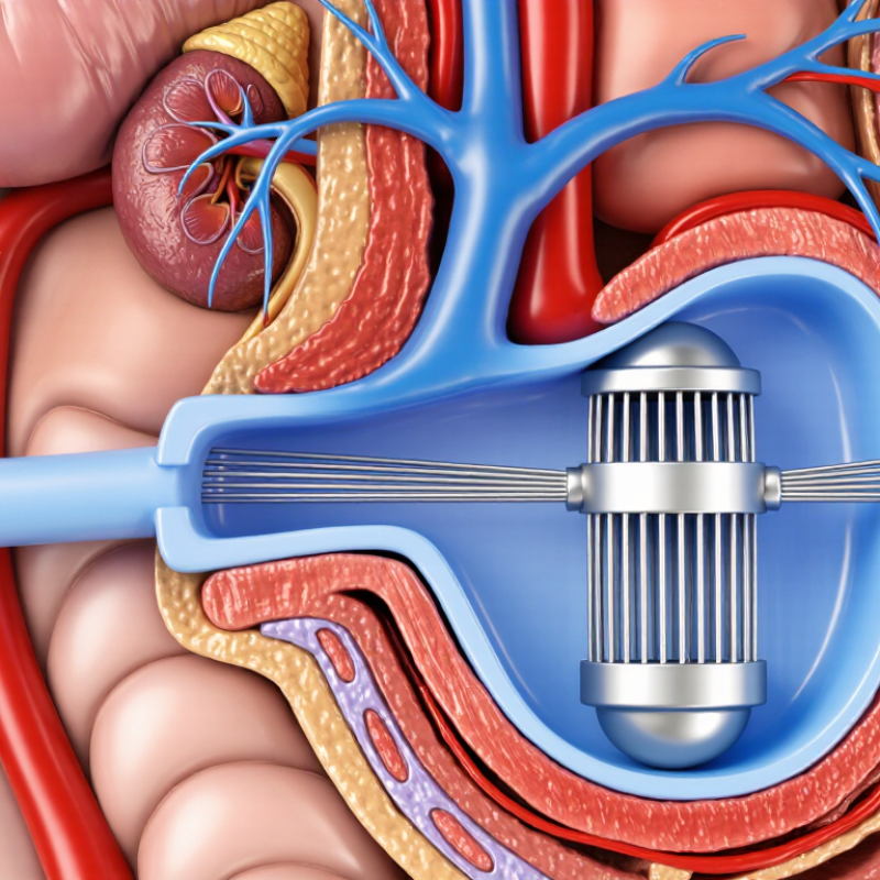
An inferior vena cava (IVC) filter is a small metal device placed inside a large abdominal vein. It can help prevent blood clots from reaching the lungs when anticoagulants are unsuitable or insufficient. Some filters are designed for later retrieval; others are permanent. Knowing which type you have is a practical first question.

The U.S. Food and Drug Administration’s 2014 safety communication discussed a decision analysis suggesting that, when a retrievable filter is no longer needed, the balance may begin to favor removal between 29 and 54 days after implantation. This is not a deadline for every patient. The Society of Interventional Radiology’s 2020 clinical practice guideline recommends a follow-up program and consideration of removal once pulmonary embolism risk has passed, unless removal risks outweigh its benefits. Timing depends on the original reason for placement, current clot risk, device position, and how long the filter has been in place.

That sounds straightforward. It often isn’t. A filter may tilt, become embedded, or develop clot around its struts, which can affect the procedure and its risks. A specialist may review imaging and medical history before recommending a plan. This guide answers the top 10 questions about vena cava filter removal, including how retrieval works, what recovery may involve, and what happens when standard removal is difficult. Bring your implant records if you have them. Small details matter. These answers support informed discussion, not a personal treatment recommendation. Your clinician can explain how the evidence applies to you.
Top 10 Questions About Vena Cava Filter Removal

What Is Vena Cava Filter Removal and Why Is It Considered?

A vena cava filter is a small device placed in the large vein that returns blood from the lower body to the heart. It can help catch blood clots traveling toward the lungs when blood-thinning medicine is unsuitable or insufficient. Removal means guiding a thin catheter through a vein, often in the neck or groin, and using imaging and specialized tools to retrieve the filter. The process is usually considered for filters designed to be temporary. Not every filter can be removed. That distinction matters.

A clinician may discuss removal when the original reason for the filter has improved, or when leaving it in place could create future concerns. These may include filter movement, damage, clot buildup, or difficulty removing it later. The decision depends on the device’s position, how long it has been in place, the patient’s health, and current clot risk.

A review may involve medical records and imaging. Removal is not risk-free; bleeding, vessel injury, or an unsuccessful attempt can occur. Sometimes keeping the filter is the safer choice.

Ask the treating specialist about the expected benefits, possible risks, and alternatives in your specific case. Details can be easy to overlook. A careful review helps.

Who May Be Eligible for Filter Removal, and When?

Who may be eligible for vena cava filter removal depends on why the filter was placed and whether that risk has changed. A person may be considered for removal when the danger of a blood clot traveling to the lungs has decreased, or when blood-thinning treatment is now suitable. The decision needs a clinician’s review.

Timing matters.

Many filters are designed to be retrievable, but some are intended to remain in place. A specialist will review the person’s medical history, current clot risk, and any symptoms. Imaging may help show the filter’s position and whether it has tilted, shifted, or become surrounded by tissue. A filter that has been in place for a long time can be more difficult to retrieve, though duration alone does not decide eligibility.

Removal is not automatically safer than leaving a filter in place. If clot risk remains high, or removal poses greater risk, continued monitoring may be appropriate. Some people also have filter-related problems, such as pain or blockage, that call for prompt assessment. Even then, removal may not be the only option. It is worth asking which risks apply personally, what testing is needed, and how soon follow-up should happen. I would not treat a scheduled appointment as proof that removal is suitable; the plan can change after evaluation.

How Is the Removal Procedure Performed?

How Is the Removal Procedure Performed?

Removal usually takes place in an interventional radiology suite, with the patient awake or lightly sedated. After reviewing the filter’s position and the reason it was placed, the clinician numbs a small area, commonly in the neck. A thin catheter passes through a vein under live X-ray guidance. A tiny snare catches the filter’s hook, then the filter is gently folded into a sheath and withdrawn. The team may inject contrast dye to check blood flow and look for injury. It is not always a quick, neat maneuver.

If the filter is tilted, embedded, or difficult to grasp, retrieval may require specialized tools or a separate approach. These details matter. Bleeding, vessel injury, or a broken filter component are possible, so the clinician weighs removal risks against the risks of leaving it in place. The Society of Interventional Radiology’s 2020 clinical practice guideline recommends follow-up and removal of retrievable filters when pulmonary embolism risk has passed, unless removal risks outweigh benefits. The FDA’s 2014 safety communication cited decision-analysis findings suggesting the balance may favor removal about 29–54 days after placement when protection is no longer needed.

Tips: Ask which vein will be accessed, what sedation is planned, and whether imaging is needed afterward. Report new chest pain, shortness of breath, or leg swelling promptly. A filter may not be removable in every case.

What Risks and Recovery Considerations Should Patients Know?

Top 10 Questions About Vena Cava Filter Removal

What Risks and Recovery Considerations Should Patients Know?

Filter removal is a procedure performed through a small incision, often in the neck or groin. A clinician guides a thin catheter to the filter using imaging. Removal may be more complex if the filter has tilted, become embedded, or trapped clot. Not every filter can be removed safely, and the decision depends on your health and imaging results. Possible complications include bleeding, infection, damage to a blood vessel, or a reaction to contrast dye. Serious complications are uncommon, but risks vary. Ask your care team which risks apply to you.

Tips: Bring a current medication list, including blood thinners. Ask whether you need to fast or adjust medicines. Tell the team about allergies and any recent bleeding.

Many patients go home the same day, but recovery instructions differ. You may have soreness or bruising near the catheter site. Keep the area clean and follow your clinician’s guidance about bathing, lifting, and activity. Contact your care team for worsening pain, swelling, fever, or bleeding that does not stop with gentle pressure. Seek urgent care for chest pain, trouble breathing, or fainting. Recovery can feel less predictable than expected; report changes rather than guessing what is normal.

Top 10 Questions About Vena Cava Filter Removal - What Risks and Recovery Considerations Should Patients Know?

Question Answer and practical considerations
1. Why might an IVC filter be removed? A retrievable inferior vena cava (IVC) filter may be considered for removal when the risk of pulmonary embolism has decreased and the filter is no longer needed. The treating clinician weighs the original reason for placement, current clot risk, and whether removal is technically appropriate.
2. How is filter removal usually performed? Removal is commonly performed by an interventional radiologist or another trained specialist using imaging guidance. A catheter is generally introduced through a vein, often in the neck or groin, and used to capture and withdraw the filter. The access site and approach depend on the individual case.
3. What are the common short-term risks? Possible risks include bruising, bleeding, pain, or a hematoma at the access site. Less commonly, infection or injury to a blood vessel may occur. The chance of complications depends on the patient’s health, medications, filter position, and procedural complexity.
4. Can removal cause serious complications? Rare but potentially serious complications include damage to the IVC or nearby structures, significant bleeding, or a filter component breaking or moving. Some filters are tilted, embedded, fractured, or surrounded by tissue, which can make retrieval more complex. The specialist should explain the risks specific to the imaging findings.
5. What if the filter cannot be removed? Some filters cannot be safely retrieved, or an attempted retrieval may be stopped if the risks are judged too high. In that situation, the clinician may recommend leaving the filter in place and arranging appropriate follow-up. A failed or deferred removal does not by itself mean an emergency.
6. Will I need imaging or tests beforehand? Before retrieval, clinicians review the filter’s position and condition, often using prior imaging and, when needed, additional imaging. Blood tests or other assessments may be appropriate based on medical history, kidney function, contrast use, and planned sedation.
7. Should I change my blood thinners before the procedure? Do not stop, start, or change anticoagulant or antiplatelet medicines without instructions from the treating team. The plan depends on why the medicine is prescribed, bleeding risk, and the procedure. Tell the team about all prescription medicines, over-the-counter medicines, and supplements.
8. What is recovery usually like? Many uncomplicated retrievals are performed as outpatient procedures, but observation or a longer stay may be needed. Patients are typically monitored after the procedure and given instructions about the access site, activity, and follow-up. Recovery time varies with the procedure and individual health.
9. What should I do at home after removal? Follow the discharge instructions, keep the access site clean and dry as directed, and avoid strenuous activity for the period specified by the care team. Ask when to resume usual activities and medicines. Contact the clinical team for worsening pain, increasing swelling, persistent bleeding, redness, or drainage at the access site.
10. Which symptoms require urgent medical attention? Seek urgent care for severe or persistent chest pain, sudden shortness of breath, fainting, coughing up blood, heavy or uncontrolled bleeding, or a rapidly enlarging access-site swelling. These symptoms can have different causes but require prompt assessment.

Important: This table provides general educational information, not personal medical advice. Whether and when to remove an IVC filter should be decided with the treating clinician, based on the patient’s circumstances and the filter’s position and condition.

What Follow-Up Care Is Needed After Removal?

After vena cava filter removal, follow-up should confirm that the access site is healing and that the original clotting risk is being managed. A clinician may check the groin or neck puncture for increasing pain, redness, swelling, or drainage. Mild bruising can happen. New shortness of breath, chest pain, or leg swelling needs urgent medical assessment. Anticoagulants are not automatic after removal; the prescriber should review why the filter was placed, clot history, bleeding risk, and current medicines.

The Society of Interventional Radiology’s 2020 guideline drew on 34 studies and recommends organized follow-up for patients with filters, including assessment for complications and retrieval when appropriate. Removal does not erase the condition that led to filter placement. Ask whether a follow-up visit or imaging is needed; testing depends on symptoms and procedural findings. The FDA’s 2014 safety communication cited a decision analysis suggesting clinicians consider removing a retrievable filter 29–54 days after placement once pulmonary-embolism risk has passed. That timing concerns removal decisions, not a fixed aftercare schedule. Recovery can feel anticlimactic. Keep the discharge instructions handy, and report symptoms rather than waiting for the next appointment.