2026 Top Achieve Biopsy Needle Types Buying Guide?

Selecting the right achieve biopsy needle requires more than comparing prices or catalog photographs. Tissue type, lesion depth, imaging method, and specimen requirements all shape the decision. A needle that performs well in a superficial liver procedure may be unsuitable for a deep, fibrous, or highly mobile target.

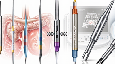
This 2026 buying guide introduces major biopsy needle types, including fine-needle aspiration, core biopsy, cutting, coaxial, and vacuum-assisted designs. It explains how gauge, shaft length, tip geometry, sample notch size, and compatibility with ultrasound, CT, or stereotactic guidance influence practical performance. Small details matter. A clear depth marker can improve control. A stable handle can reduce hand fatigue during a lengthy procedure. A coaxial system may also limit repeated skin punctures when multiple samples are needed.

Clinical experience remains essential. Published evidence, manufacturer instructions, sterilization information, and quality certifications should be reviewed before purchase. Product claims deserve careful checking against real workflow requirements. Marketing language can sound impressive, but it cannot replace validated performance data. Fit matters.

No single needle suits every patient or facility. Budget limits, staff training, local protocols, and maintenance support may change the final choice. This guide cannot remove every judgment call, and some comparisons remain difficult without hands-on evaluation. A responsible buyer should request samples, inspect packaging and safety features, and consult qualified clinicians or procurement specialists. The best selection balances diagnostic yield, patient safety, operator confidence, and dependable supply. That balance is practical, not perfect.
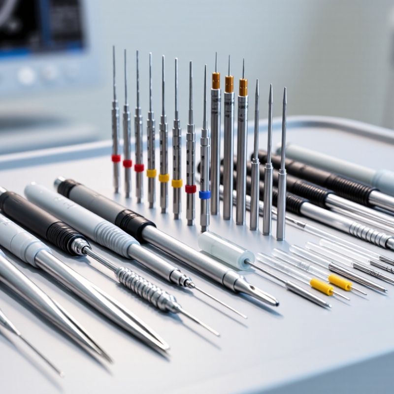
2026 Top Achieve Biopsy Needle Types Buying Guide?

Biopsy Needle Types and Their Core Design Features

Biopsy Needle Types and Their Core Design Features

Biopsy needle selection begins with the tissue target, not the catalog page. Fine-needle aspiration needles use narrow cannulas for cytology and usually reduce tissue disruption. Core biopsy needles remove intact cylinders, preserving architecture for histological assessment. Vacuum-assisted designs collect larger or multiple specimens through a rotating aperture. No needle is universally best.

Gauge, bevel geometry, cutting edge, throw length, and stylet design strongly influence performance. A sharper outer cannula can improve cutting efficiency, while a longer throw may capture a larger core. Coaxial systems can reduce repeated tissue punctures during image-guided procedures. Echogenic markings also matter when the needle tip must remain visible on ultrasound. Small details matter.

The International Agency for Research on Cancer reported about 20 million new cancer cases worldwide in 2022. That burden increases demand for accurate tissue diagnosis, but market growth should not replace clinical judgment. A 2024 MarketsandMarkets report projected continued expansion in the global biopsy devices market, supported by image-guided procedures and minimally invasive diagnosis. Exact forecasts vary between reports.

In practice, buyers should compare specimen quality, deployment control, imaging compatibility, sterilization status, and documented clinical evidence. A thinner needle may reduce resistance, yet it can yield less tissue. A larger needle may improve architecture, but handling becomes more demanding. That trade-off is easy to overlook. Product specifications alone cannot predict every patient outcome. According to the College of American Pathologists, specimen adequacy and pathology workflow remain central to reliable diagnosis.

How Each Biopsy Needle Type Works

Biopsy needle choice depends on the tissue, lesion size, and diagnostic question. Fine-needle aspiration uses a thin needle to collect loose cells. It works well for cytology, but it may provide limited tissue structure. Core needles use a cutting tip and a spring-loaded mechanism. They remove small tissue cylinders, preserving architecture for histological examination. Vacuum-assisted needles use suction to capture multiple larger samples through one access point.

Each type works differently inside the body. A fine needle creates minimal disruption. A core needle advances, cuts, and withdraws a tissue core. A vacuum-assisted system pulls tissue into the sampling chamber before cutting it. The operator must control depth, angle, and specimen handling carefully. A larger needle is not automatically better. It may improve tissue volume, yet increase bleeding risk or patient discomfort. That trade-off deserves honest discussion.

Tips: Match the needle gauge to the target and imaging method. Confirm the sampling path before insertion. Prepare enough passes for laboratory needs. Label every specimen immediately. Review image quality after each pass. Do not rely on habit alone. Local protocols and specialist judgment remain essential. No needle performs perfectly in every case, and even experienced teams should reassess unclear results or inadequate samples.

Clinical Uses and Tissue Sampling Applications

2026 Top Achieve Biopsy Needle Types Buying Guide?

Clinical use should guide every biopsy needle choice. Fine-needle aspiration needles collect individual cells for cytology. They are useful for thyroid nodules, lymph nodes, and selected soft-tissue lesions. The sample is small. Precision matters.

Core biopsy needles remove narrow tissue cylinders for histological review. They can preserve tissue architecture, helping specialists assess tumors, fibrosis, and inflammatory changes. Spring-loaded designs may support consistent sampling, while coaxial systems can reduce repeated skin punctures during image-guided procedures. Larger needles may provide more tissue, but they can also increase bleeding risk and patient discomfort.

Vacuum-assisted needles collect multiple cores through one access point. They are often considered for breast lesions and selected image-guided applications. Needle length, gauge, cutting mechanism, and compatibility with ultrasound, CT, or stereotactic guidance should match the target’s depth and location. Difficult anatomy changes the decision.

A technically successful procedure can still produce a poor diagnosis if the specimen is fragmented, too small, or placed in the wrong medium. Pathology requirements should be confirmed before sampling. Patient history, anticoagulant use, lesion vascularity, and operator experience also influence safety. There is no universal “best” needle. In practice, the ideal choice is sometimes a compromise between tissue quality, access, bleeding control, and patient comfort. That compromise deserves review.

Key Factors for Comparing Biopsy Needles in 2026

Top Achieve Biopsy Needle Types Buying Guide

Key Factors for Comparing Biopsy Needles in 2026

Choosing a biopsy needle starts with the tissue target, not the product label. Fine-needle aspiration suits cellular sampling, while core needles preserve larger tissue structures. Vacuum-assisted systems may collect multiple cores through one access point. Each type creates different handling demands. Match the needle to the lesion, imaging method, and required pathology.

Gauge affects both tissue volume and insertion resistance. A wider needle may provide stronger core architecture, but it can increase discomfort and bleeding risk. Length should reach the target without excessive deflection. Tip geometry also matters. Cutting edges, side ports, and bevel angles influence penetration and sample quality. Under ultrasound or CT guidance, visibility and needle stiffness deserve careful comparison.

Reliable purchasing includes more than specifications. Review sterility assurance, packaging integrity, compatibility with introducers, and clear instructions for use. Check whether the handle supports controlled advancement with gloved hands. A smooth trigger can reduce sudden movement. Ask for performance data, not only marketing claims. Independent evaluations and clinician feedback can reveal weaknesses that brochures miss. Cost per procedure should include wastage, reprocessing limits, and staff training. The cheapest option may become expensive after poor samples or repeat procedures. No needle is ideal. Real-world testing may expose small problems, such as a slippery grip or inconsistent core length. That is why a documented trial with trained professionals remains valuable.

2026 Top Achieve Biopsy Needle Types Buying Guide? - Key Factors for Comparing Biopsy Needles in 2026

Biopsy Needle Type Typical Gauge Range Sample Type Best-Suited Clinical Use Specimen Architecture Sampling Characteristics Key Advantages Important Limitations Imaging or Guidance Compatibility Primary Buying Factors
Fine-Needle Aspiration (FNA) Needle 22–25G Cellular aspirate and fluid Cytology of superficial masses, thyroid nodules, lymph nodes, and selected deep lesions Limited; cells are generally obtained without preserved tissue structure Usually rapid and minimally invasive; diagnostic yield depends strongly on lesion characteristics and sampling technique Small puncture, relatively low tissue trauma, and efficient fluid or cellular sampling May be inadequate for tissue architecture, grading, receptor testing, or some lymphoma workups; repeat sampling may be required Ultrasound, endoscopic ultrasound, CT, and palpation-guided procedures Tip visibility, bevel sharpness, needle stiffness, aspiration control, compatibility with cytology accessories, and accurate gauge labeling
Core Biopsy Needle 14–20G Small cylindrical tissue core Breast, liver, kidney, prostate, soft-tissue, and other solid-organ lesions Good preservation of tissue architecture when the core is adequate and properly handled Collects one or more tissue cores; sample quality varies with needle diameter, lesion location, targeting, and number of passes Supports histology, immunohistochemistry, and many molecular or biomarker assessments More invasive than FNA; bleeding, pain, and insufficient-sample risk remain procedure-dependent Ultrasound, stereotactic mammography, CT, MRI-compatible systems, and other image-guided approaches Core length, cutting efficiency, firing mechanism, penetration depth, coaxial compatibility, sample retention, and deployment control
Coaxial Core Biopsy System Introducer commonly 17–19G; inner biopsy needle commonly 14–20G Multiple tissue cores through a single access route Deep or difficult-to-access lesions where several samples may be needed Usually good, because the inner core needle can be passed repeatedly through the introducer Allows repeated sampling without creating a new skin puncture for every pass May reduce repeated access trauma and help maintain a consistent trajectory Requires sufficient working space and careful management of introducer position; added components increase setup complexity Commonly used with CT, ultrasound, and other image-guided interventions Introducer rigidity, depth markings, inner-needle fit, locking mechanism, visibility, obturator design, and sterile configuration
Vacuum-Assisted Biopsy (VAB) Probe Approximately 7–13G Multiple larger tissue fragments or cores Breast lesions, microcalcifications, sampling of small lesions, and selected image-guided procedures Generally strong tissue architecture, although specimen quality depends on fragmentation and handling Vacuum draws tissue into a side aperture for sequential sampling, often allowing multiple specimens through one insertion High sampling volume, useful for clustered targets, and may reduce the need for repeated needle insertion Larger access channel, higher equipment and procedural complexity, and potential for increased bruising or hematoma Most commonly stereotactic or ultrasound-guided; system-specific imaging compatibility should be confirmed Aperture size, vacuum consistency, cutting speed, specimen chamber capacity, tissue fragmentation, noise, ergonomics, and system compatibility
End-Cutting or Franseen-Style Core Needle Commonly 19–25G, depending on application Core-like tissue fragments Selected endoscopic ultrasound-guided sampling of pancreatic, gastrointestinal, liver, and other deep lesions Often better than conventional aspiration for obtaining histologic fragments, but results vary by lesion and technique Cutting edges or a specialized tip capture tissue during forward advancement or controlled sampling Can provide tissue fragments with relatively small needle diameters and may support histologic assessment Yield can be affected by fibrosis, necrosis, lesion mobility, operator technique, and specimen-processing method Endoscopic ultrasound and other specialized image-guided procedures Tip geometry, tissue capture, flexibility, echogenicity, stylet performance, compatibility with the endoscope, and reprocessing or single-use requirements
Bone Marrow Aspiration Needle Commonly 15–18G Liquid marrow aspirate Cytology, flow cytometry, cellularity assessment, and selected hematologic investigations Limited in the aspirate alone; architecture is assessed more reliably with a concurrent trephine core Withdraws marrow through a stylet-equipped needle after cortical entry Efficient collection of marrow particles and aspirate for laboratory analysis Aspirate may be diluted, dry, or inadequate; does not replace an intact core when architecture is required Usually landmark-guided; image guidance may be used in selected patients or difficult anatomy Stylet security, handle ergonomics, depth control, cortical penetration, aspiration connection, and ease of cleaning or disposal
Bone Marrow Trephine Biopsy Needle Commonly 8–11G Solid cylindrical bone marrow core Marrow architecture, fibrosis assessment, cellularity, infiltrative disease, and staging investigations Designed to preserve a longer, more representative marrow core than aspiration alone Obtains an intact core after controlled advancement and rotation through the cortical bone Provides structural information that cannot be obtained from aspirate alone More painful and technically demanding than aspiration; core fragmentation or crush artifact can affect interpretation Usually landmark-guided, with imaging assistance in selected circumstances Core retention, cutting edge, depth markings, handle control, stylet fit, resistance during advancement, and specimen integrity
Soft-Tissue or Musculoskeletal Core Needle Commonly 14–18G Soft-tissue core specimen Muscle, tendon-adjacent masses, subcutaneous lesions, and selected sarcoma evaluations Generally suitable for histology when sufficient viable tissue is obtained Designed to obtain tissue from firm, mobile, or heterogeneous soft-tissue targets Supports architectural assessment and ancillary laboratory testing Sampling error is possible in heterogeneous lesions; proximity to nerves, vessels, or functional structures requires careful planning Ultrasound, CT, and MRI-planned or MRI-compatible procedures where applicable Tip sharpness, throw length, flexibility, tissue retention, coaxial access, and compatibility with the planned imaging modality
General comparison for procurement and procedure planning. Gauge ranges, needle construction, sample yield, and imaging compatibility vary by intended application and device configuration; clinical selection should follow local protocols and qualified clinician judgment.
2026 comparison priorities: match the needle to the required specimen type, target depth and consistency, imaging method, desired number of passes, tissue-processing workflow, patient safety requirements, and total procedural cost—not gauge alone.

Safety, Compatibility, and Purchasing Considerations

2026 Top Achieve Biopsy Needle Types Buying Guide

Safety, Compatibility, and Purchasing Considerations

Choosing a biopsy needle starts with the procedure, not the catalogue image. Core needles collect tissue cylinders, while fine-needle aspiration needles obtain cells or fluid. Vacuum-assisted systems may gather larger samples during selected image-guided procedures. Each design creates different handling and bleeding considerations. Clinicians should match needle gauge, length, tip geometry, and tissue path to the planned imaging method. A needle that is too short may force awkward positioning. A needle that is too stiff may reduce control near delicate structures. Small details matter.

Compatibility deserves careful checking. Confirm the needle works with the biopsy gun, coaxial introducer, ultrasound guide, or computed tomography workflow. Verify connection standards, firing depth, marker visibility, and sterile packaging. The instructions for use should define compatible accessories and patient-use limits. Do not rely on a familiar appearance. Similar devices can have different fittings. That mistake is easy to make.

Purchasing teams should request validation documents, lot traceability, shelf-life data, and complaint-response procedures. Review published clinical evidence, but remember that study results may not transfer to every operator or lesion. Training records matter as much as unit price. A lower-cost needle can increase repeat passes, procedure time, or discarded specimens. That is an uncomfortable calculation. Ask for sample evaluation when permitted, then assess specimen quality, deployment control, packaging integrity, and staff feedback. Local regulatory requirements and facility policies must guide the final order.

2026 Top Achieve Biopsy Needle Types Buying Guide: Safety, Compatibility, and Purchasing Considerations

Needle gauge indicates the approximate outer diameter: a lower gauge number represents a larger needle. The chart shows representative midpoints from commonly specified clinical gauge ranges; the final choice must follow the procedure, anatomy, imaging method, patient condition, and the device manufacturer’s instructions for use.

Safety

Confirm sterility, single-use status, tissue-targeting requirements, bleeding risk, and imaging guidance before purchase. Larger needles may obtain more tissue but can increase procedural trauma.

Compatibility

Check compatibility with the biopsy gun or vacuum system, introducer, coaxial sheath, imaging table, and sampling accessories. Verify length, tip design, firing depth, and connection type.

Purchasing

Compare gauge range, usable length, specimen quality, packaging, shelf life, documentation, training requirements, and availability rather than selecting by gauge alone.