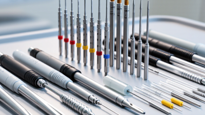
How to Choose a Tru Cut Biopsy Needle in 2026
Choosing a tru cut biopsy needle in 2026 is not a matter of picking the sharpest-looking device. The right choice depends on the target, tissue type, imaging guidance, access route, and clinician experience. A small breast lesion, a firm lymph node, and a deep soft-tissue mass may call for different needle lengths, gauges, and firing mechanisms. Details matter. A clear ultrasound view, a steady hand, and a needle suited to the target can support consistent core samples while helping clinicians plan a careful procedure.
This guide compares practical selection factors, including sample quality, control, compatibility, handling, and device documentation. It also highlights questions to raise with a qualified clinician or supplier, such as whether the needle fits the planned imaging approach and whether its instructions are clear. No single model is best for every case. The checklist helps, but it cannot replace clinical judgment. And real-world choices are sometimes messier than product tables suggest.
No source was provided for a verifiable quotation from a named tru cut biopsy needle expert, so I have not invented one. A reliable attributed quote should be added only after checking its original source.

Define the Target and Review ACR Image-Guided Biopsy Criteria
Choosing a Tru-Cut biopsy needle starts with defining the target, not picking a gauge. Identify the lesion’s size, depth, location, and tissue type on current imaging. Note nearby vessels, pleura, bowel, or other structures that could affect the access route. A small, mobile target may require a different approach from a deep, firm mass. Be specific.
Review the current ACR practice parameter for the relevant organ and imaging method. These parameters address appropriate image guidance, patient preparation, consent, procedural safety, and specimen handling. They support consistent practice, but do not replace clinical judgment or local protocols. Confirm that the planned route is visible and that the patient can maintain the needed position. Check relevant bleeding risks and medication history according to clinical policy. Small details matter.
Then match needle length and gauge to the target and planned trajectory. A longer needle may be needed for a deep lesion; a thicker core may provide more tissue but can require greater care near sensitive structures. The best choice depends on the clinical question and operator experience. Confirm how many cores are needed with the pathology team when possible. Recheck the target just before sampling. Imaging can be imperfect, and I have seen a seemingly clear route look less convincing once the patient is positioned. When uncertain, pause and reassess rather than forcing the original plan.
Compare Common 14G–20G Tru-Cut Needle Gauges and Core Sizes
A Tru-Cut needle’s gauge describes its outer diameter: the lower the gauge number, the thicker the needle. Approximate outer diameters are 2.1 mm for 14G, 1.65 mm for 16G, 1.27 mm for 18G, and 0.91 mm for 20G. The tissue core is usually narrower than the needle. Its actual width and length also depend on the cutting notch and device design, so gauge alone cannot predict the specimen size precisely.
A 14G needle generally collects a larger, more substantial core, which may help when tissue architecture matters. It also creates a larger needle tract. A 16G offers a middle ground, while 18G is commonly selected when a smaller access path is useful. A 20G can be appropriate when a finer needle is needed, but the resulting core may provide less tissue for analysis. These are tendencies, not guarantees. Notch lengths vary.
The right choice depends on the target’s size, depth, tissue type, imaging guidance, and the pathologist’s sample requirements. For a small lesion, a thicker needle may be harder to position accurately. For a firm or heterogeneous target, a very fine core may be less representative. Ask the clinical team what specimen dimensions their assessment requires, and confirm the needle’s notch length and validated core specifications. Numbers help. They do not replace judgment.
Match Throw Length and Sampling Notch to Target Depth
Choosing a Tru Cut biopsy needle means matching its throw length to the target’s depth and size.
Measure the planned path on imaging, then account for the distance from the needle tip to the sampling notch. The notch must sit within the lesion when the device fires; a long throw can pass beyond a small target or approach nearby structures.
Small targets need restraint. A shorter throw may offer better control, while a longer notch can collect more tissue when the lesion safely accommodates it.
Confirm the device’s actual specifications, since usable notch length and total throw are not interchangeable.
A 2018 systematic review and meta-analysis in BMC Cancer reported pooled sensitivity of about 97% and specificity of about 99% for ultrasound-guided breast core-needle biopsy.
Those figures describe diagnostic performance across studies, not a guarantee for any needle or target. Image quality, lesion location, operator technique, and specimen adequacy also matter.
A neat rule—“deeper target, longer throw”—sounds practical, but can mislead when the target is narrow or close to a vessel.
Recheck the intended notch position in the imaging plane, and follow local clinical protocols. Sometimes the safest choice is not obvious.
Capture enough tissue without letting the sampling window extend beyond the intended target.
Verify FDA 510(k) Status and Applicable Sterilization Standards
Before selecting a Tru Cut biopsy needle, verify its exact model in the FDA 510(k) database. A 510(k) clearance applies to a specific device and intended use; it is not the same as FDA approval. Match the listing to the needle’s labeling, including its gauge, length, and whether it is supplied sterile. The FDA’s MDUFA IV performance goals set a 90% target for traditional 510(k) decisions within 90 FDA days. That review timeline is not evidence of clinical superiority or sterility. Check the exact model. If the supplier’s documents do not match the database entry, pause and request clarification.
Next, confirm the sterilization method and applicable standards on the label and supporting documentation. Ethylene oxide sterilization is covered by ISO 11135; radiation sterilization by ISO 11137; and moist-heat sterilization by ISO 17665. Sterile barrier packaging requirements are addressed by ISO 11607. Check the current editions and FDA-recognized standards before relying on a certificate. A sterile symbol alone does not identify the process, its validation, or the package’s integrity. Look for a clear method, lot traceability, and an intact pouch with no punctures or moisture. FDA’s 510(k) database and its recognized consensus standards database provide useful verification records, but neither replaces review of the device’s labeling. Documents can be incomplete. That deserves attention.
How to Choose a Tru Cut Biopsy Needle in 2026: Verify FDA 510(k) Status and Applicable Sterilization Standards
How to use this checklist: The bars count verification points in this guide, not product ratings or clearance rates. Match the exact needle model and intended use to its FDA 510(k) record. Confirm the stated sterilization method, validation, and labeling; standards to check may include ISO 11135 for ethylene oxide, ISO 11137 for radiation, ISO 17665 for moist heat, and ISO 11737 for microbiological testing. Confirm the applicable editions and requirements for the specific device and market.
Compare Organ-Specific Diagnostic Yield and Complication Data
Choosing a Tru-Cut biopsy needle in 2026 means matching the device to the organ and the clinical question. Diagnostic yield is not a needle specification alone: tissue quality, lesion size, imaging guidance, and pathology handling all matter. Small details matter. Compare studies that define adequacy similarly, rather than treating every reported “yield” as equivalent.
For liver lesions, a core may preserve tissue architecture for diagnosis, while bleeding risk depends on patient factors, lesion location, and the route taken. In kidney biopsy, adequate cortical tissue is essential; bleeding remains a key complication to weigh against the expected diagnostic benefit. For lung lesions, pneumothorax risk is strongly influenced by the access path and surrounding lung, not just needle gauge. These are patterns, not promises. Reported complication rates vary across populations and study methods.
A larger core can provide more tissue, but may require a different balance of control and risk in delicate or vascular targets. Review organ-specific data for sample adequacy, repeat-biopsy rates, and complications requiring treatment—not just any recorded adverse event. Also check whether results apply to the patient’s anatomy and imaging approach. The evidence is not perfectly clean; comparisons can be difficult. Discuss needle choice and precautions with the treating team, using current local protocols and the individual patient’s risk profile.
How to Choose a Tru-Cut Biopsy Needle in 2026: Organ-Specific Diagnostic Yield and Complication Data
A practical comparison of commonly sampled organs. Reported ranges are approximate and vary with imaging guidance, lesion characteristics, needle gauge, operator experience, and how each study defines a diagnostic result.
| Organ / Target | Typical Diagnostic Performance | Key Complication Data | Needle and Sampling Considerations | Guidance and Risk Checks | Practical Selection Point |
|---|---|---|---|---|---|
| Liver | Image-guided core biopsy commonly obtains tissue adequate for diagnosis in about 90–98% of cases in published series. Adequacy depends on the indication, lesion visibility, and tissue-processing requirements. | Major bleeding is uncommon, generally reported at under 1% in many contemporary series; pain and minor bleeding are more frequent. Risk is higher with coagulopathy, thrombocytopenia, or certain lesion locations. | Choose a core length and gauge that provide enough tissue for the requested histology and ancillary testing while accounting for the target and the safest trajectory. A coaxial approach may reduce repeated capsular punctures. | Ultrasound or CT guidance is selected according to target visibility and access. Review coagulation status, platelet count, medications affecting hemostasis, and the planned route. | For a focal lesion, prioritize accurate targeting and a safe path; for diffuse disease, follow the clinician’s protocol for representative parenchymal sampling. |
| Kidney | Native-kidney biopsy is diagnostic in approximately 90–95% of cases in many series when adequate glomerular tissue is obtained. Pathology may require material for light microscopy, immunofluorescence, and electron microscopy. | Bleeding is the principal complication. Published series commonly report transfusion-requiring bleeding around 1–2%; less severe hematomas are more frequent. Rates differ by patient risk and definitions used. | Use a needle and sampling plan that provide adequate cortex for the laboratory’s required studies. Avoid compromising tissue adequacy solely to reduce gauge without considering the clinical protocol. | Real-time ultrasound guidance is commonly used. Assess blood pressure, hemoglobin, platelet count, coagulation status, kidney anatomy, and medications that affect bleeding risk. | Confirm specimen allocation with pathology before the procedure; diagnostic success depends on appropriate tissue handling as well as needle choice. |
| Lung | CT-guided percutaneous core biopsy often has diagnostic accuracy in the approximate 85–95% range, with results influenced by lesion size, location, and the proportion of viable tissue sampled. | Pneumothorax is commonly reported in roughly 15–25% of procedures; about 4–6% require chest-tube drainage in many pooled reports. Rates vary substantially with technique and patient factors. | Needle gauge, throw length, and coaxial technique should be selected around lesion size, pleural distance, and the need for tissue architecture. Minimize traversed aerated lung when feasible. | CT guidance is commonly used. Consider emphysema, lesion depth, fissure or bulla traversal, respiratory status, and the safest patient position and route. | Balance tissue needs against pneumothorax risk; a small or deep lesion may require a more tailored trajectory and sampling plan. |
| Breast | Image-guided core needle biopsy has high diagnostic performance; reported sensitivity is generally above 95% in many settings. Imaging–pathology concordance is essential and may prompt repeat sampling when results do not explain the imaging finding. | Bruising, hematoma, and short-term soreness can occur. Clinically significant bleeding or infection is uncommon; complication rates depend on lesion location, technique, and the definitions used in a study. | Select a device and sampling approach suitable for lesion depth, imaging modality, and the amount of tissue needed. Multiple representative cores are commonly obtained under the local protocol. | Ultrasound, stereotactic, or MRI guidance may be used depending on how the lesion is best seen. Review antithrombotic medication and planned compression or aftercare. | Prioritize reliable targeting and radiologic–pathologic concordance; if the sample is inadequate or discordant, further evaluation may be needed. |
Related Posts
-
2026 Top Achieve Biopsy Needle Types Buying Guide?
-
7 Best Biopsy Needles to Achieve Accurate Sampling
-
China Top Tru Cut Biopsy Needle Manufacturers Suppliers
-
How to Choose the Right Bone Biopsy Needle for Accurate Diagnosis?
-
How to Choose the Right Bone Biopsy Needle for Your Needs?
-
How to Choose the Right Bone Biopsy Needle for Your Medical Needs